Contact Information
University of Illinois
260 MRL, MC-230
600 South Mathews Avenue
Urbana, IL 61801
Research Areas
Biography
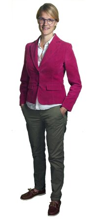
Professor van der Veen obtained her B.S. and M.S. degrees in Chemistry from the Swiss Federal Institute of Technology (ETH) in Zürich in 2006. In 2010 she received her Ph.D. in the field of ultrafast X-ray science from the École Polytechnique Fédérale de Lausanne (EPFL) and the Swiss Light Source. After working as a postdoctoral fellow at the California Institute of Technology, she became a project group leader at the Max Planck Institute for Biophysical Chemistry in Göttingen. She joined the University of Illinois faculty as an Assistant Professor in April 2015, and is affiliated with the Frederick Seitz Materials Research Laboratory and the Materials Science and Engineering Department. She is interested in the study of atomic-scale mechanisms of light-induced processes, such as photoswitching, photovoltaics and photocatalysis.
Research Interests
excited-state structural dynamics of photoswitching, photocatalytic and photovoltaic materials; ultrafast spectromicroscopy; ultrafast electron microscopy; ultrafast X-ray science
Research Description
The conversion of light energy into chemical energy is a topic of uttermost importance in today's world. Among the general goals is to develop materials that can efficiently convert and store energy from the sun, or nanomaterials that can be rapidly switched between two states allowing them to be used in data storage devices. A fundamental understanding of the photophysical processes involved in light-energy conversion is prerequisite for developing such new materials with desired properties.
Within this context, our research focuses on several types of light-sensitive (molecular) materials: (i) switchable metal-organic complexes, (ii) functional nanomaterials relevant to photocatalysis, and (iii) photovoltaic perovskite materials. We would like to understand how the energy contained in a photon is channeled onto pathways and into states that can be used for specific functions. How fast and efficient are excited-state relaxation processes and how do electronic and structural degrees of freedom couple?
To tackle these questions, we employ a variety of ultrafast spectroscopic and microscopic methods with time resolutions ranging from femtoseconds (fs) to nanoseconds (ns).
(1) Ultrafast Optical Spectroscopy and Microscopy
Optical spectroscopy in the UV-visible spectral range is mainly sensitive to the occupation and energy of valence orbitals close to the Fermi level. Transient absorption spectroscopy with ~100 fs time resolution is used to follow the energy relaxation processes upon excitation into well-defined absorption bands originating from metal-to-ligand charge-transfer (MLCT), metal- or ligand-centered transitions. By interfacing transient absorption spectroscopy with a laser-scanning confocal microscope, we will study relaxation processes in nanoscale materials, such as nanoparticles and thin films.
(2) Ultrafast X-ray Spectroscopy at Large-Scale Facilities
The transformation of spectroscopic observables in the UV-visible range into bond distances requires detailed knowledge about the potential energy surfaces of the states involved. While this is clear for small molecules, it becomes more ambiguous when the system grows in complexity and size, e.g. when solute-solvent interactions come into play. In order to overcome these difficulties, we use probing methods based on high-energy radiation (X-rays) or particles (electrons) to probe the systems under study. The high energy entails a short wavelength which is used to obtain the desired atomic-spatial resolution, without the necessity of a priori knowledge about potential energy surfaces.
X-ray absorption spectroscopy (XAS) is particularly appealing for the study of (metal-organic) molecular materials, as it is a local probe of both the electronic and geometric structure, and it can be implemented in any medium. In our lab, we use short (fs-ps) X-ray pulses from synchrotron radiation facilities or X-ray free-electron lasers (XFEL) to probe the dynamics after excitation with a fs laser pulse. Dedicated computer codes are employed to simulate and fit the X-ray spectra in order to extract the excited-state dynamics.
(3) Ultrafast Electron Microscopy
We exploit the high spatial and temporal resolutions of ultrafast electron microscopy (UEM) to reveal the electronic and structural dynamics of nanostructured materials. Compared to ultrafast optical and X-ray methods, UEM exhibits a superior spatial imaging resolution (sub-nm), and it offers the possibility to characterize morphology (via imaging), geometric structure (via diffraction), and electronic structure (via spectroscopy, EELS) of materials - all within the same table-top setup.
UEM is now becoming an increasingly attractive tool that combines the atomic-scale spatial resolution of conventional TEM, with the high temporal (fs-ns) resolution of optical spectroscopy. The principle of UEM is based on the generation of ultrashort electron pulses using the photoelectric effect at the cathode. Reversible excited-state processes can be studied in stroboscopic mode at high repetition rates, while irreversible processes are probed by single-shot and movie-mode detection.
The UEM machine that is used in our lab is currently being developed at the Frederick Seitz Materials Research Laboratory in collaboration with the group of Prof. Jian-Min Zuo.
Awards and Honors
Sofja Kovalevskaja Award of the Alexander von Humboldt Foundation (April 2014)
Independent Max Planck Group Leader position appointed by the President of the Max Planck Society (March 2014)
Special mention in the EPFL Doctorate Award election (April 2012)
Prospective Researcher Fellowship of the Swiss National Science Foundation (April 2010)
Swiss Chemical Society prize for the best poster talk in Physical Chemistry, SCS Fall Meeting, Lausanne (Sept. 2007)
Doctoral fellowship awarded by the Doctoral School of Photonics, EPFL (Nov. 2006)
Courses Taught
CHEM 590 X
CHEM 442
Additional Campus Affiliations
Assistant Professor, Materials Science and Engineering
Assistant Professor, Materials Research Lab
Recent Publications
Zandi, O., Sykes, A., Cornelius, R., Alcorn, F., & Van Der Veen, R. (Accepted/In press). Periodic Lensing from a Photoemitted Electron Gas Undergoing Cyclotron Oscillations. Microscopy and Microanalysis, [181]. https://doi.org/10.1017/S1431927620014671
Zandi, O., Sykes, A. E., Cornelius, R. D., Alcorn, F. M., Zerbe, B. S., Duxbury, P. M., Reed, B. W., & van der Veen, R. M. (2020). Transient lensing from a photoemitted electron gas imaged by ultrafast electron microscopy. Nature communications, 11(1), [3001]. https://doi.org/10.1038/s41467-020-16746-z
Pomarico, E., Kim, Y. J., García De Abajo, F. J., Kwon, O. H., Carbone, F., & Van Der Veen, R. M. (2018). Ultrafast electron energy-loss spectroscopy in transmission electron microscopy. MRS Bulletin, 43(7), 497-503. https://doi.org/10.1557/mrs.2018.148
Park, S. T., & van der Veen, R. M. (2017). Modeling nonequilibrium dynamics of phase transitions at the nanoscale: Application to spin-crossover. Structural Dynamics, 4(4), [044028]. https://doi.org/10.1063/1.4985058
Vanacore, G. M., Van Der Veen, R. M., & Zewail, A. H. (2015). Origin of axial and radial expansions in carbon nanotubes revealed by ultrafast diffraction and spectroscopy. ACS Nano, 9(2), 1721-1729. https://doi.org/10.1021/nn506524c