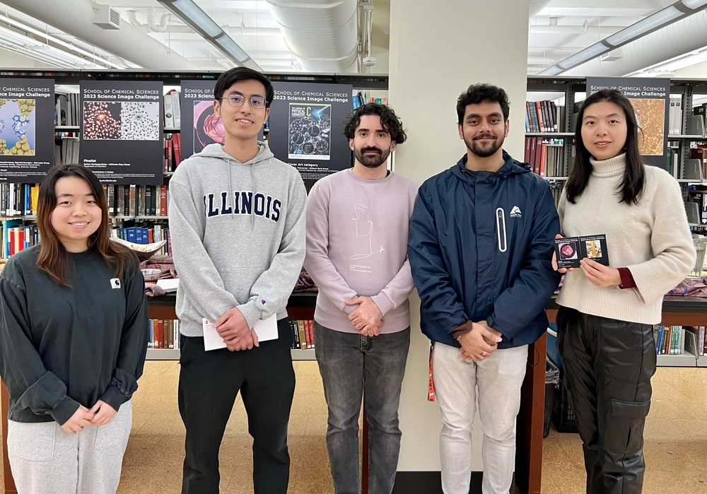
The School of Chemical Sciences has announced the 2023 Science Image Challenge list of winners and finalists, featuring images highlighting research projects by graduate students, postdoctoral researchers, and faculty members in the Department of Chemistry and Chemical and Biomolecular Engineering.
The winners and finalists were celebrated during an awards ceremony on March 7, 2024, in the Chemistry Library in Noyes Laboratory.
The annual SCS Image Challenge highlights computer-assisted or traditional scientific images that feature an object, process or technique within a scientific research project in the departments of chemistry and chemical and biomolecular engineering at Illinois. The challenge is open to SCS graduate and undergraduate students, postdoctoral associates and fellows, and staff. The entrant’s Principal Investigator must be a faculty member or an affiliate/adjunct of the departments of chemistry or chemical and biomolecular engineering.
The winning images and finalists' images from the 2023 Science Image Challenge will go on exhibit at Willard Airport in Champaign. The School of Chemical Sciences, the Department of Chemistry and the Department of Chemical and Biomolecular Engineering thank Willard Airport for hosting the winning images exhibit.
Main Category Winner
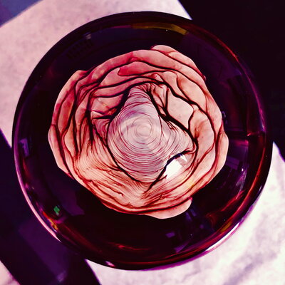
By Xiaolin Liu, Prof. Jeffrey Moore Lab, Department of Chemistry
Title: Rose in the flask
Description: Some roses grow from flowerbeds, and others appear in chemistry labs. This rose-like hue and pattern formed at the bottom of a flask as dichloromethane solvent evaporated from an aromatic compound sample. The polycyclic aromatic hydrocarbons show useful electronic properties and also imparts color.
Cover Art Category Winner
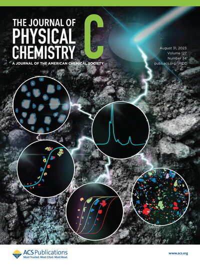
By Yating Feng & Krishna C. Polavaram, Garg Lab, Departments of Civil & Environmental Engineering and Department of Chemistry
Description: Using Raman imaging, for the first time, we report phase-specific particle size distributions and shape characteristics for each phase in a set of 10 commercially available cements. By integrating these physical characteristics with chemical abundance, we establish a new parameter (CSQ) that can predict the 72 h cumulative heat of hydration (R2 = 0.86). This finding underscores the significance of phase-specific particle size distributions in predicting the performance of cementitious systems.
Main Category finalists
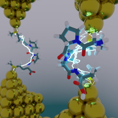
By Moeen Meigooni, Emad Tajkhorshid Lab, Center for Biophysics and Quantitative Biology, Department of Biochemistry, Department of Chemistry, Beckman Institute
Title: Do Peptides Dream of Electric Sheep?
Description: An atomistic model of a molecular break junction: an oligopeptide is caught between two gold electrodes, forming a single-molecule circuit. Using a combined experimental computational approach, we show that secondary structure plays an important role in determining electron transport in peptides. Image rendered using VMD and Blender.
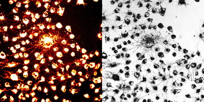
By Suritra Bandyopadhyay, Jeff Chan Lab, Department of Chemistry
Title: Driving a muscle
Description: What might resemble spiders or fireworks are, in fact, exquisite crystals of formazan. Astonishingly, these patterns are crafted not by 3D printing but by living cells. The reducing nature of mitochondrial enzymes yields insoluble Formazan crystals from 3-(4,5-dimethylthiazol[1]2-yl)-2,5-diphenyltetrazolium bromide, thereby used to assess the cellular toxicity of chemicals.

By Xiaolin Liu, Jeffrey Moore lab, Department of Chemistry
Title: Golden Necklace
Description: The “golden necklace” pattern was formed because of trapped air and water pockets during frontal ring-opening metathesis polymerization, an energy-efficient technique for fabricating high-quality thermosets and composites used in engineering applications.
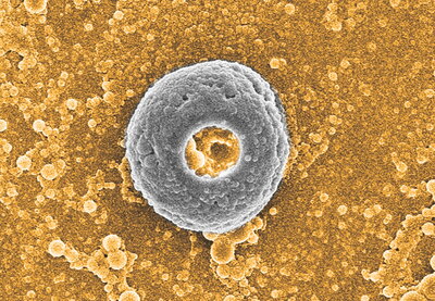
By Wangsuk Oh, Xiao Su lab, Chemical and Biomolecular Engineering
Title: Ugly Duckling Poly Donuts
Description: Polymer electrolyte complexes can form particles with diverse shapes like bread dough. The electron microscopy image shows an uncommon donut-shaped (or toroidal) particle surrounded by spherical nanoparticles formed when drying the polymer electrolyte mixtures.
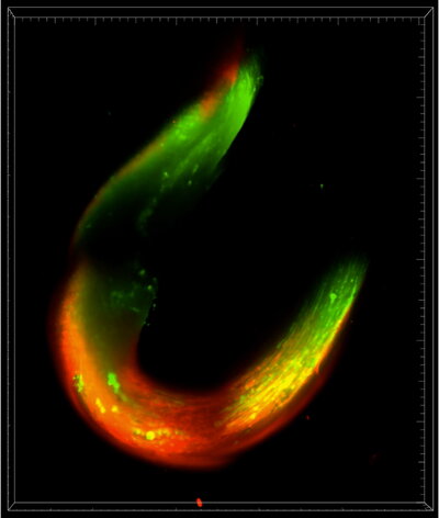
By Kai-Yu Huang, Kong lab, Department of Chemical & Biomolecular Engineering.
Untitled
Description: The image represented a 3D muscle ring made by C2C12 skeletal muscle. The cells were stained with fluorescent dye to label mature myotubes which showing in green color. This image is acquired with the lightsheet microscope in IGB.